Reception: ![]() +36/30 show
+36/30 show
6721 Szeged, Osztrovszky utca 12.
Open every week-day since 1996
Open on week-days from 8 AM to 9 PM
24 578 satisfied customers
Serving you since 1996
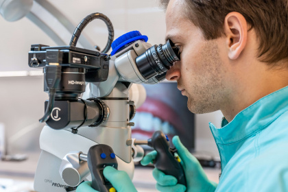
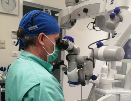
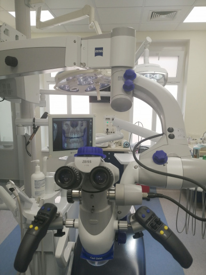
When doing dental treatments, we often use a magnifying glass, since magnification is indispensable for the precise execution of the upcoming treatments. Teeth themselves, just like their building components and the tissues surrounding them are complex structures. At Dentha Ultramodern Dental and Implantology Clinic, our dentists – using Zeiss Opmi ProErgo microscopes, that have been representing for decades the highest peak in the sector of dental technology – can execute the dental treatments with a convincing accuracy. This also means, that more precision and much better treatment results are achievable (however, it is noticeable, that in case of root canal treatments, no guarantee can be given in this respect, due to the anatomic structure of teeth).
With our top-graded Zeiss OPMI ProErgo equipments, we can achieve even a 30 times magnification. This helps to protect your healthy teeth saving by these significant costs on the long run:
In case of the following interventions, our top graded-dental microscope is of great use:
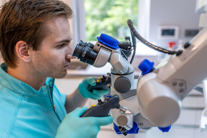
In case of teeth with multiple root canal, it is very unlikely to find the one or two extra roots by a simple eye inspection. The microscope helps us to clean sufficiently the nest of the bacteria and to fill up the shelters.
If you wish to preserve your root canal tooth the longest possible, we recommend our high-tech microscope root canal treatment! The advantages of root canal treatments executed by Zeiss OPMI ProErgo equipment:
It may occur, that the inflammation of root canal, or the inflammation around the root-top is so complex, that we can not save the tooth by the root canal treatment. In this case we may need to eliminate the root-top or in the worst case, pull out a tooth. The implant treatment following the resection and the tooth extraction can also be done by microscope.
vissza a kérdésekhez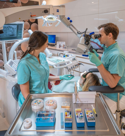
The microscopic root canal treatments in our clinic are executed by Dr. Kristóf Benedek, who is an expert in saving multi-root teeth and providing other micro-surgery treatments.
He has acquired his solid competence from a few Hungarian and foreign doctors (for example: Dr. Judit Jeney, Dr. Glenn Van As, Dr. Domenico Massironi, Dr. Alessandro Conti, Dr. Antonio Chaniotis), whose theoretical and practical hands-on courses he both accomplished.
vissza a kérdésekhez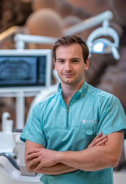
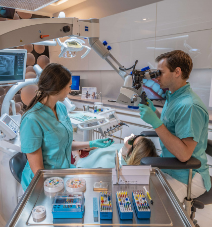
By using our special dental microscope, we can accomplish the root canal treatment with an incomparably much higher efficiency and precision in comparison with the former procedures.
In most of the cases, there is no way for the dentist to inspect within the sinus roots of the tooth, therefore he can only count on his experience and anticipation.
The magnification makes it possible to find an easier solution in those cases that turn out to be extremely difficult (curved, invisible, narrowed roots) because of the individual anatomy of teeth. For example, the upper molar teeth contain 4 dental root canals in 94% of the cases, and these can only and exclusively be detected by magnification.
By microscope, the inspection becomes possible even down to the very bottom of the root canal, thus, the canal can be properly cleaned. Cracks, that have a significant impact on the solvability of tooth, also become perceptible by microscope.
Generally, in such a case, instead of root canal treatment, tooth extraction is the right choice, by doing this, un unsuccessful root canal treatment is also evitable. Using our top-graded, high-definition Zeiss microscope, we ended up with the „blind-flight based” treatments, consequently, we could push over 90% the rate of the successful treatments.
vissza a kérdésekhez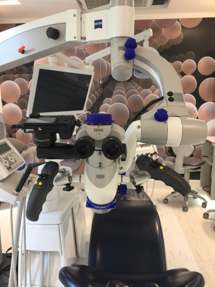
Root canal treatment is an intervention aiming to save the tooth. During the root canal treatment executed by the microscope, the dentist, after removing the decay-zone, opens the root canal and by that can reach the infected tissues.
The removal of infected tissues and the cleaning of root canal system goes along with manually used flexible microtools specially developed for microscope treatment and with mechanical tools and disinfection solutions.
Depending on the infection-degree of the root canal system, at the end of the treatment, the dentist provides the tooth with a temporary root filling containing anti-inflammatory substance, that is to be finalised a forthcoming occasion. In case of a weaker inflammation, the dentist prepares the definite root filling.
During the repeated treatment of teeth that were previously exposed to some unsuccessful root canal treatment, the dentist removes the former root filling by applying a chemical solution and mechanically, with the help of an ultra flexible root-treating tool. Following this, he disinfects the root canal by using the procedure described just above, to stop the spread of damaging processes.
When he is done with it, depending on the case, immediately or during the forthcoming visits, the dentist provides the tooth with a new filling, contributing by this to save it.
Nevertheless, the success rate of professionally accomplished root canal treatments is extremely high, in root canal treatments, just as in any other medical intervention a 100% success guarantee can not be given.
Written by: Dr. Kristóf Benedek
vissza a kérdésekhez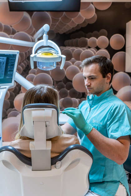
What kind of oral surgery interventions can be executed by microscope?
Just like in case of the root canal treatment, one major advantage of the microscopic oral surgery interventions is the 30× magnification which helps to accomplish the tooth extraction, the resection, and the implants with a maximal accuracy. The other important advantage is that this intervention touches the effectively treatable zone with the highest possible safety and doesn’t damage the healthy parts at all. This can be especially important when for example at the extraction of wisdom tooth, the tooth stretches into the root canal. Without the microscope, we would assume this operation only in clinical circumstances, but when using a microscope, the patient in need of such an intervention doesn’t need to go to the hospital.
Who provides the microscopic oral surgery interventions at Dentha Clinic?
The complicated microscopic tooth extraction, the wisdom-tooth and extra tooth extractions, the operational extraction of impacted tooth and the resection are all executed by Dr. Gábor Benedek and Dr. Kristóf Benedek.
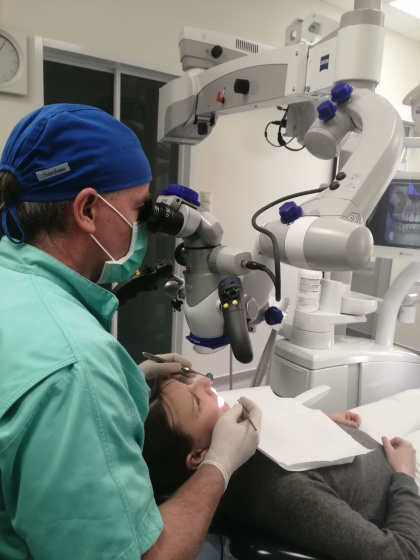
By microscope tooth filling, inlay-onlay-overlay fillings, veneer, crown and bridge can also be done. Regarding the longevity of fillings and crowns, the edge-closing is an important factor, since this decides how easy it would be to clean the surface of the tooth, and how is for bacteria to settle down. In comparison with treatments by simple eye-inspection, when making fillings and crowns by microscope we can reach a much higher level of accuracy in fitting, we can increase their longevity and prevent the occurrence of decay zones as well -providing a sufficient oral hygienic, and a regular dental hygienic control.
The other advantage of the microscopic aesthetic interventions is that the dentist can prepare the tooth for the crown with a significantly less healthy material loss and with an increased accuracy. In case of a tooth burring preceding the filling, the dentist should take away much less from the healthy tooth material and the cementing (sticking) material can be used also with an increased accuracy.
In our dentistry, we work with the most modern filling materials. If necessary, we prepare temporary dentures and fillings so that, if possible, you never have to be toothless.
vissza a kérdésekhez